
Many people become concerned when they notice raised bumps or nodules at the back of their tongue. In most cases, these structures are normal anatomical features known as papillae that support taste perception, digestion, and immune defense. However, changes in appearance, sensation, or persistence can signal irritation, infection, or nutritional imbalance. Understanding the biological role of tongue structures and maintaining effective hygiene practices helps prevent unnecessary anxiety while promoting oral wellness. This article explains tongue anatomy, warning indicators, and evidence-based care strategies that support both oral and systemic health.
The surface of the tongue is not smooth by design. It contains specialized structures called papillae, which perform critical biological functions.
• Hosting taste receptors
• Assisting with food manipulation and swallowing
• Supporting oral immune surveillance
• Helping maintain microbial balance
Circumvallate Papillae
Large dome-shaped bumps arranged in a V-shape at the back of the tongue (typically 8–12). These are the bumps most people notice and are usually normal.
Fungiform Papillae
Smaller mushroom-shaped structures near the tip and sides, containing taste buds.
Foliate Papillae
Located along the rear sides; often more visible in younger individuals.
Filiform Papillae
Most abundant type; provides texture and grip for food but does not contain taste buds.
Seeing bumps at the back of the tongue is therefore typically a sign of healthy anatomy rather than disease.
Although papillae are normal, certain changes should be monitored.
• Mechanical irritation from hot or spicy foods
• Minor trauma from biting
• Temporary inflammation
• Persistent redness or swelling
• White or yellow coating
• Painful nodules
• Bleeding or ulceration
• Changes lasting more than two weeks
These symptoms may reflect:
• Oral infections
• Nutritional deficiencies
• Microbial imbalance
• Chronic irritation
Professional evaluation is recommended when abnormalities persist.
The tongue hosts a significant portion of oral bacteria and contributes to the overall oral microbiome.
• Reduces odor-causing bacteria
• Supports microbial balance
• Improves taste perception
• Lowers plaque transfer to teeth
• Gently brush or scrape the tongue daily
• Rinse after meals
• Maintain hydration
• Use antimicrobial rinses when appropriate
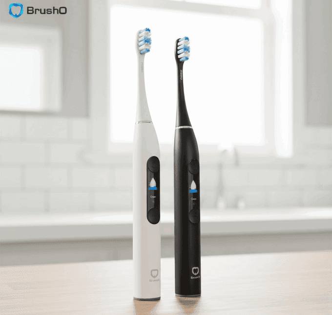
Advanced oral care tools such as BrushO assist in maintaining comprehensive cleaning by guiding full-mouth coverage beyond just tooth surfaces.
Oral tissues often reflect broader physiological conditions.
• White coating — immune stress or microbial imbalance
• Smooth red tongue — vitamin deficiency signals
• Swollen papillae — inflammatory response
• Persistent lesions — require clinical assessment
The tongue functions as an early biological indicator, highlighting the importance of consistent monitoring.
Bumps at the back of the tongue are typically normal papillae essential for taste, digestion, and immune defense. Awareness of anatomical variation prevents unnecessary concern, while understanding warning signs supports early intervention when needed. Integrating proper tongue hygiene into daily oral care routines contributes to both oral microbiome balance and overall wellness. Smart oral care technologies like BrushO help ensure comprehensive cleaning coverage, supporting healthier habits and improved long-term oral outcomes.
Feb 20
Feb 19

Squamous cell carcinoma is not a single, uniform disease, and two of its oral variants illustrate the extremes of its behavior. Verrucous carcinoma is a slow-growing, exophytic tumor that almost never metastasizes, while spindle cell carcinoma is an aggressive, sarcomatoid malignancy that can be ...

Most people expect a toothache to be the first sign of pulp disease, but some forms of pulpitis are surprisingly silent. A pulp polyp is the visible result of chronic hyperplastic pulpitis, a condition in which the dental pulp grows out of a large carious opening as a fleshy mass. Recognizing thi...

Dry mouth is not merely uncomfortable; it is a medical condition with real consequences for teeth, speech, swallowing and quality of life. When the salivary glands cannot produce enough saliva, medications called sialogogues can restore some of the missing fluid. Pilocarpine and cevimeline are th...

A blister in the mouth can be the first warning of a systemic autoimmune disease, and two disorders deserve particular attention in the dental chair: pemphigus vulgaris and mucous membrane pemphigoid. Both produce chronic oral ulceration, yet their immunologic targets, clinical behavior and long-...

When infection drains from a tooth, the body often creates a visible escape route, and the dentist must interpret these fistulous openings correctly. A parulis is the common gum boil that signals chronic periapical infection, while an oroantral fistula is a communication between the mouth and the...

Among the fibro-osseous lesions of the jaws, the ossifying fibroma holds a unique position because it is a true neoplasm, unlike the reactive cemento-osseous dysplasia discussed in an earlier article. It grows continuously, expands bone and requires surgical removal, and its cemental variant adds...

Not all keratocystic lesions of the jaw behave alike, and one of the most important distinctions in oral pathology separates the orthokeratinized odontogenic cyst from its better-known relative, the odontogenic keratocyst. Although the two lesions look similar under the microscope at first glance...

Periodontal disease is usually a slowly progressive infection, but a rare form called necrotizing ulcerative periodontitis destroys tissue with alarming speed. Unlike ordinary periodontitis, this condition produces necrotic, cratered papillae, deep pocketing and rapid bone loss, and it appears al...

Every parent has met the child who refuses to eat because of painful mouth sores, and most of these children have one of two viral illnesses: herpangina or hand, foot and mouth disease. Both are caused by enteroviruses, both produce fever and oral ulcers, and both are overwhelmingly common in you...

Cementoosseous dysplasia is one of the most common fibro-osseous lesions found on dental radiographs, yet it is also one of the most frequently misdiagnosed. The lesion replaces normal bone with cellular fibrous tissue and cementum-like material, and it appears in several forms that range from a ...

Build an oral health data network quickly and at low cost in a way that everyone can build.


Address: RM 1427, 14/F RADIO CITY 505, HENNESSY RD CAUSEWAY BAY, HONG KONG
Code: 000000
Phone: +852-53758652
Email: customerservice@brusho.com