
Plaque accumulation begins within hours after brushing, forming a biofilm that adheres tightly to tooth surfaces and gumlines. Many people assume that occasional intensive cleaning sessions can compensate for daily inconsistencies, yet dental science consistently shows the opposite: early plaque removal is far more effective at preventing oral disease than reactive deep cleaning. Once plaque matures and mineralizes into tartar, it becomes resistant to routine brushing and requires professional intervention. Preventive biofilm disruption maintains microbial balance, protects enamel integrity, and stabilizes gum health. This article explores why timing, frequency, and precision in plaque control outweigh aggressive cleaning efforts and how smart brushing technologies help manage plaque before it evolves into pathology.
Dental plaque is a living microbial biofilm composed of:
• Bacterial colonies
• Salivary proteins
• Food particles
• Extracellular structural matrices
It begins forming within 4–12 hours after cleaning.
Early-stage plaque is:
• Soft
• Loosely attached
• Easily removable
Delayed removal allows plaque to:
• Thicken
• Harden
• Increase bacterial complexity
• Trigger inflammation
Timing determines whether plaque remains manageable or becomes pathogenic.
Removing plaque before maturation prevents:
• Matrix reinforcement
• Microbial communication networks
• Acidogenic species dominance
Early disruption stops disease processes before activation.
Plaque bacteria metabolize sugars and release acids that:
• Demineralize enamel
• Increase sensitivity
• Initiate cavities
Frequent plaque removal shortens acid exposure cycles and preserves tooth mineral density.
Accumulated plaque along the gumline causes:
• Gingival irritation
• Bleeding
• Immune activation
Consistent early cleaning stabilizes soft tissue defenses and prevents periodontal progression.
After 24–72 hours:
• Plaque begins calcifying
• Becomes tartar
• Adheres strongly to enamel
At this stage:
• Brushing is ineffective
• Professional scaling required
Mature plaque hosts more pathogenic species associated with:
• Periodontitis
• Tissue destruction
• Bone loss
Reactive cleaning addresses damage — not root cause.
Attempting aggressive removal later may cause:
• Gum trauma
• Enamel abrasion
• Sensitivity
Preventive care avoids excessive mechanical stress.
Common hygiene patterns that delay plaque removal:
• Skipping nighttime brushing
• Rushing sessions
• Missing molars or gumline
• Inconsistent routine timing
These behaviors allow plaque to mature repeatedly. Small daily gaps accumulate into long-term clinical issues.
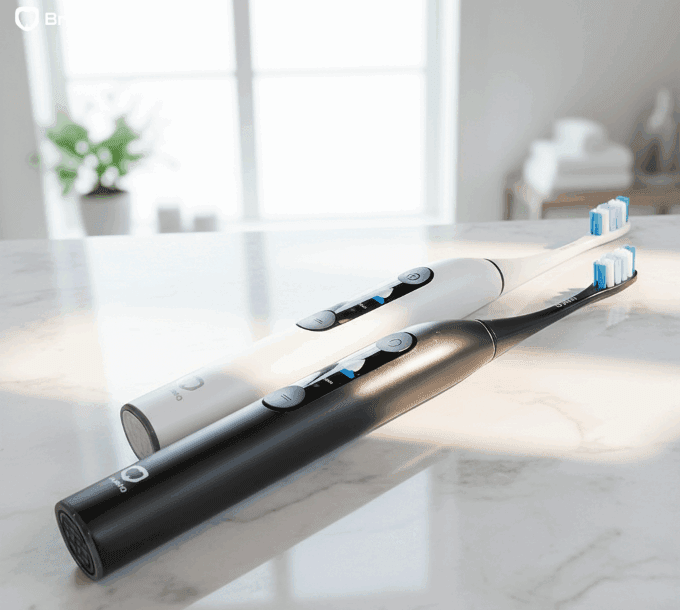
Precision brushing tools like BrushO improve preventive removal by addressing habit variability.
Ensures no surface becomes a persistent plaque reservoir.
Maintains effective cleaning without tissue damage.
Identifies neglected regions before biofilm stabilizes.
Habit tracking encourages consistent timing and frequency.
Early intervention becomes measurable and repeatable rather than guesswork.
Managing biofilm adaptation helps:
• Lower cavity incidence
• Prevent gingival inflammation
• Maintain microbiome diversity
• Reduce tartar formation
• Stabilize enamel integrity
Oral health improves when the hygiene strategy evolves faster than microbial adaptation.
Oral biofilm is a responsive biological system shaped by daily hygiene behavior. Rather than simply accumulating, microbial communities reorganize and adapt to brushing consistency, technique, and environmental conditions. Understanding this dynamic interaction reframes oral hygiene as ecological management rather than routine cleaning. AI-guided brushing technologies support this approach by minimizing predictable gaps and improving coverage precision. By influencing how biofilm evolves, individuals gain proactive control over long-term oral health outcomes.

Squamous cell carcinoma is not a single, uniform disease, and two of its oral variants illustrate the extremes of its behavior. Verrucous carcinoma is a slow-growing, exophytic tumor that almost never metastasizes, while spindle cell carcinoma is an aggressive, sarcomatoid malignancy that can be ...

Most people expect a toothache to be the first sign of pulp disease, but some forms of pulpitis are surprisingly silent. A pulp polyp is the visible result of chronic hyperplastic pulpitis, a condition in which the dental pulp grows out of a large carious opening as a fleshy mass. Recognizing thi...

Dry mouth is not merely uncomfortable; it is a medical condition with real consequences for teeth, speech, swallowing and quality of life. When the salivary glands cannot produce enough saliva, medications called sialogogues can restore some of the missing fluid. Pilocarpine and cevimeline are th...

A blister in the mouth can be the first warning of a systemic autoimmune disease, and two disorders deserve particular attention in the dental chair: pemphigus vulgaris and mucous membrane pemphigoid. Both produce chronic oral ulceration, yet their immunologic targets, clinical behavior and long-...

When infection drains from a tooth, the body often creates a visible escape route, and the dentist must interpret these fistulous openings correctly. A parulis is the common gum boil that signals chronic periapical infection, while an oroantral fistula is a communication between the mouth and the...

Among the fibro-osseous lesions of the jaws, the ossifying fibroma holds a unique position because it is a true neoplasm, unlike the reactive cemento-osseous dysplasia discussed in an earlier article. It grows continuously, expands bone and requires surgical removal, and its cemental variant adds...

Not all keratocystic lesions of the jaw behave alike, and one of the most important distinctions in oral pathology separates the orthokeratinized odontogenic cyst from its better-known relative, the odontogenic keratocyst. Although the two lesions look similar under the microscope at first glance...

Periodontal disease is usually a slowly progressive infection, but a rare form called necrotizing ulcerative periodontitis destroys tissue with alarming speed. Unlike ordinary periodontitis, this condition produces necrotic, cratered papillae, deep pocketing and rapid bone loss, and it appears al...

Every parent has met the child who refuses to eat because of painful mouth sores, and most of these children have one of two viral illnesses: herpangina or hand, foot and mouth disease. Both are caused by enteroviruses, both produce fever and oral ulcers, and both are overwhelmingly common in you...

Cementoosseous dysplasia is one of the most common fibro-osseous lesions found on dental radiographs, yet it is also one of the most frequently misdiagnosed. The lesion replaces normal bone with cellular fibrous tissue and cementum-like material, and it appears in several forms that range from a ...

Build an oral health data network quickly and at low cost in a way that everyone can build.


Address: RM 1427, 14/F RADIO CITY 505, HENNESSY RD CAUSEWAY BAY, HONG KONG
Code: 000000
Phone: +852-53758652
Email: customerservice@brusho.com