
Many people become concerned when they notice raised bumps or nodules at the back of their tongue. In most cases, these structures are normal anatomical features known as papillae that support taste perception, digestion, and immune defense. However, changes in appearance, sensation, or persistence can signal irritation, infection, or nutritional imbalance. Understanding the biological role of tongue structures and maintaining effective hygiene practices helps prevent unnecessary anxiety while promoting oral wellness. This article explains tongue anatomy, warning indicators, and evidence-based care strategies that support both oral and systemic health.
The surface of the tongue is not smooth by design. It contains specialized structures called papillae, which perform critical biological functions.
• Hosting taste receptors
• Assisting with food manipulation and swallowing
• Supporting oral immune surveillance
• Helping maintain microbial balance
Circumvallate Papillae
Large dome-shaped bumps arranged in a V-shape at the back of the tongue (typically 8–12). These are the bumps most people notice and are usually normal.
Fungiform Papillae
Smaller mushroom-shaped structures near the tip and sides, containing taste buds.
Foliate Papillae
Located along the rear sides; often more visible in younger individuals.
Filiform Papillae
Most abundant type; provides texture and grip for food but does not contain taste buds.
Seeing bumps at the back of the tongue is therefore typically a sign of healthy anatomy rather than disease.
Although papillae are normal, certain changes should be monitored.
• Mechanical irritation from hot or spicy foods
• Minor trauma from biting
• Temporary inflammation
• Persistent redness or swelling
• White or yellow coating
• Painful nodules
• Bleeding or ulceration
• Changes lasting more than two weeks
These symptoms may reflect:
• Oral infections
• Nutritional deficiencies
• Microbial imbalance
• Chronic irritation
Professional evaluation is recommended when abnormalities persist.
The tongue hosts a significant portion of oral bacteria and contributes to the overall oral microbiome.
• Reduces odor-causing bacteria
• Supports microbial balance
• Improves taste perception
• Lowers plaque transfer to teeth
• Gently brush or scrape the tongue daily
• Rinse after meals
• Maintain hydration
• Use antimicrobial rinses when appropriate
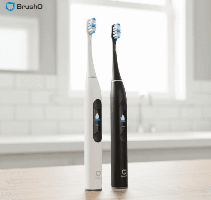
Advanced oral care tools such as BrushO assist in maintaining comprehensive cleaning by guiding full-mouth coverage beyond just tooth surfaces.
Oral tissues often reflect broader physiological conditions.
• White coating — immune stress or microbial imbalance
• Smooth red tongue — vitamin deficiency signals
• Swollen papillae — inflammatory response
• Persistent lesions — require clinical assessment
The tongue functions as an early biological indicator, highlighting the importance of consistent monitoring.
Bumps at the back of the tongue are typically normal papillae essential for taste, digestion, and immune defense. Awareness of anatomical variation prevents unnecessary concern, while understanding warning signs supports early intervention when needed. Integrating proper tongue hygiene into daily oral care routines contributes to both oral microbiome balance and overall wellness. Smart oral care technologies like BrushO help ensure comprehensive cleaning coverage, supporting healthier habits and improved long-term oral outcomes.
Feb 20
Feb 19

Hyperdontia is the presence of one or more teeth in excess of the normal dental formula, and the resulting teeth are described as supernumerary. The condition is common enough to appear in routine practice but variable enough in presentation that the decision to intervene is rarely straightforwar...

Few patients complete orthodontic treatment without asking whether their teeth will move back. The honest answer is that they will, to some degree, in the majority of cases. Relapse is not a failure of treatment but a predictable biological response, and understanding why it happens is the basis ...

The mandible is the largest and most frequently fractured bone of the facial skeleton, and its management sits at the interface between trauma surgery and restorative dentistry. Treatment has shifted over four decades from prolonged maxillomandibular fixation towards rigid internal fixation with ...

Chronic liver disease affects hundreds of millions of people worldwide, and its prevalence continues to rise with the epidemic of metabolic liver disease. The liver is central to haemostasis, drug metabolism and immune competence, and its failure therefore alters the risk profile of almost every ...

Excessive gingival display, commonly described as a gummy smile, is one of the most frequent aesthetic concerns presented to dental practitioners. The condition is not a single entity but a clinical presentation that can arise from skeletal, gingival, muscular or dental factors, and the treatment...

Drug-induced gingival overgrowth is a well-recognised adverse effect of several widely prescribed medications, and it remains one of the most common drug-related findings in periodontal practice. The condition produces firm, fibrous enlargement of the gingiva that can interfere with speech, masti...

Denture stomatitis is an inflammatory condition of the denture-bearing mucosa that affects a large proportion of removable prosthesis wearers, yet it is frequently dismissed as a cosmetic annoyance. The condition is driven by a Candida biofilm on the fitting surface of the prosthesis, and it is m...

Chronic kidney disease affects an estimated 850 million people worldwide, and the number of patients surviving on dialysis or with a renal transplant continues to rise. These patients carry an oral disease burden that is substantially higher than that of the general population, and their medical ...

Angular cheilitis is an inflammatory lesion of the labial commissures that presents with erythema, fissuring and crusting at the corners of the mouth. It is one of the most common conditions encountered in general dental practice, yet it is frequently treated as a simple fungal infection and allo...

Adverse reactions to dental materials are uncommon but they are not rare, and they are frequently misdiagnosed because the oral mucosa responds to allergens in a limited number of ways. A burning sensation, a persistent ulcer or an unexplained area of erythema may all represent a hypersensitivity...

Construire rapidement et à faible coût un réseau de données sur la santé bucco-dentaire de manière que tout le monde puisse y contribuer.
Construire rapidement et à faible coût un réseau de données sur la santé bucco-dentaire de manière que tout le monde puisse y contribuer.


Address: RM 1427, 14/F RADIO CITY 505, HENNESSY RD CAUSEWAY BAY, HONG KONG
Code: 000000
Phone: +852-53758652
Email: customerservice@brusho.com