
Think your toothbrush head can last a year? Think again. Using the same brush head for too long can seriously compromise your dental hygiene—and possibly your health. Worn bristles can’t clean properly, harbor bacteria, and may even damage your gums. In this article, we’ll explain why regular brush head replacement is essential, how often you really need to change it, and how BrushO’s free brush head program makes it easier than ever to stay protected and save money.
Over time, bristles lose their shape and stiffness, which reduces their ability to remove plaque and debris. A brush head that looks “fine” may have already lost 30%–40% of its cleaning effectiveness.
Old brush heads can become breeding grounds for millions of bacteria. The moist environment, combined with leftover toothpaste and mouth bacteria, creates the perfect space for microbial growth—even mold in some cases.
Frayed bristles not only clean less effectively, but they can also irritate your gums and wear down tooth enamel. If you’re brushing twice daily with a worn head, you might be hurting your teeth more than helping.
According to the American Dental Association (ADA), brush heads should be replaced every 3 months. You should also replace it sooner if:
The bristles look worn or splayed
You’ve recently been sick (e.g., flu, cold, strep throat)
The head has been dropped or exposed to contamination
🦷 Pro tip: With BrushO, your smart toothbrush reminds you when it’s time to replace the brush head—so you’ll never forget again.
Let’s be honest—it’s easy to forget. Most of us don’t mark a calendar or set reminders. Some assume that unless the bristles are obviously damaged, it’s fine. But even if your brush head looks okay, its microscopic wear can already be putting your teeth at risk.
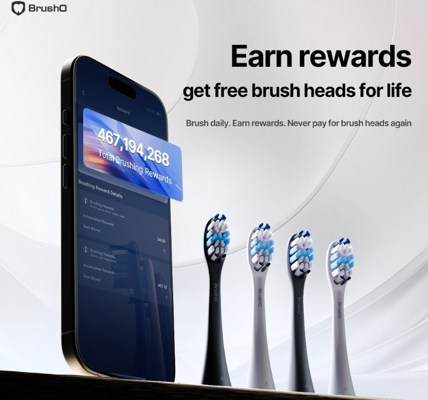
BrushO solves this problem with an industry-disrupting model: you get free brush heads for life.
Use BrushO to brush your teeth daily
Earn points for each brushing session via the BrushO app
Redeem points to claim free replacement brush heads
This means you’re rewarded for good habits—and never need to worry about overspending on essential care.
Fresh bristles remove bacteria and plaque more effectively, leading to fresher breath and less buildup.
New brush heads are gentle on the gumline and better at reducing inflammation and preventing gingivitis.
BrushO’s smart system syncs better with fresh bristles, giving you more accurate feedback and reports.
Bristles are frayed or bent
Discoloration (bristles turning yellow or gray)
Unusual smell or sticky residue
Your app alerts you (if using a smart brush like BrushO)
It’s been over 3 months
Replacing your toothbrush head isn’t just a hygiene tip—it’s a non-negotiable for good oral care. And with BrushO’s AI reminders and lifetime brush head plan, keeping your teeth protected has never been easier—or more affordable.
You’re not just brushing smarter—you’re brushing better, longer, and healthier.

Squamous cell carcinoma is not a single, uniform disease, and two of its oral variants illustrate the extremes of its behavior. Verrucous carcinoma is a slow-growing, exophytic tumor that almost never metastasizes, while spindle cell carcinoma is an aggressive, sarcomatoid malignancy that can be ...

Most people expect a toothache to be the first sign of pulp disease, but some forms of pulpitis are surprisingly silent. A pulp polyp is the visible result of chronic hyperplastic pulpitis, a condition in which the dental pulp grows out of a large carious opening as a fleshy mass. Recognizing thi...

Dry mouth is not merely uncomfortable; it is a medical condition with real consequences for teeth, speech, swallowing and quality of life. When the salivary glands cannot produce enough saliva, medications called sialogogues can restore some of the missing fluid. Pilocarpine and cevimeline are th...

A blister in the mouth can be the first warning of a systemic autoimmune disease, and two disorders deserve particular attention in the dental chair: pemphigus vulgaris and mucous membrane pemphigoid. Both produce chronic oral ulceration, yet their immunologic targets, clinical behavior and long-...

When infection drains from a tooth, the body often creates a visible escape route, and the dentist must interpret these fistulous openings correctly. A parulis is the common gum boil that signals chronic periapical infection, while an oroantral fistula is a communication between the mouth and the...

Among the fibro-osseous lesions of the jaws, the ossifying fibroma holds a unique position because it is a true neoplasm, unlike the reactive cemento-osseous dysplasia discussed in an earlier article. It grows continuously, expands bone and requires surgical removal, and its cemental variant adds...

Not all keratocystic lesions of the jaw behave alike, and one of the most important distinctions in oral pathology separates the orthokeratinized odontogenic cyst from its better-known relative, the odontogenic keratocyst. Although the two lesions look similar under the microscope at first glance...

Periodontal disease is usually a slowly progressive infection, but a rare form called necrotizing ulcerative periodontitis destroys tissue with alarming speed. Unlike ordinary periodontitis, this condition produces necrotic, cratered papillae, deep pocketing and rapid bone loss, and it appears al...

Every parent has met the child who refuses to eat because of painful mouth sores, and most of these children have one of two viral illnesses: herpangina or hand, foot and mouth disease. Both are caused by enteroviruses, both produce fever and oral ulcers, and both are overwhelmingly common in you...

Cementoosseous dysplasia is one of the most common fibro-osseous lesions found on dental radiographs, yet it is also one of the most frequently misdiagnosed. The lesion replaces normal bone with cellular fibrous tissue and cementum-like material, and it appears in several forms that range from a ...

Costruire rapidamente e a basso costo una rete di dati sulla salute orale in modo che tutti possano creare.
Costruire rapidamente e a basso costo una rete di dati sulla salute orale in modo che tutti possano creare.


Address: RM 1427, 14/F RADIO CITY 505, HENNESSY RD CAUSEWAY BAY, HONG KONG
Code: 000000
Phone: +852-53758652
Email: customerservice@brusho.com