
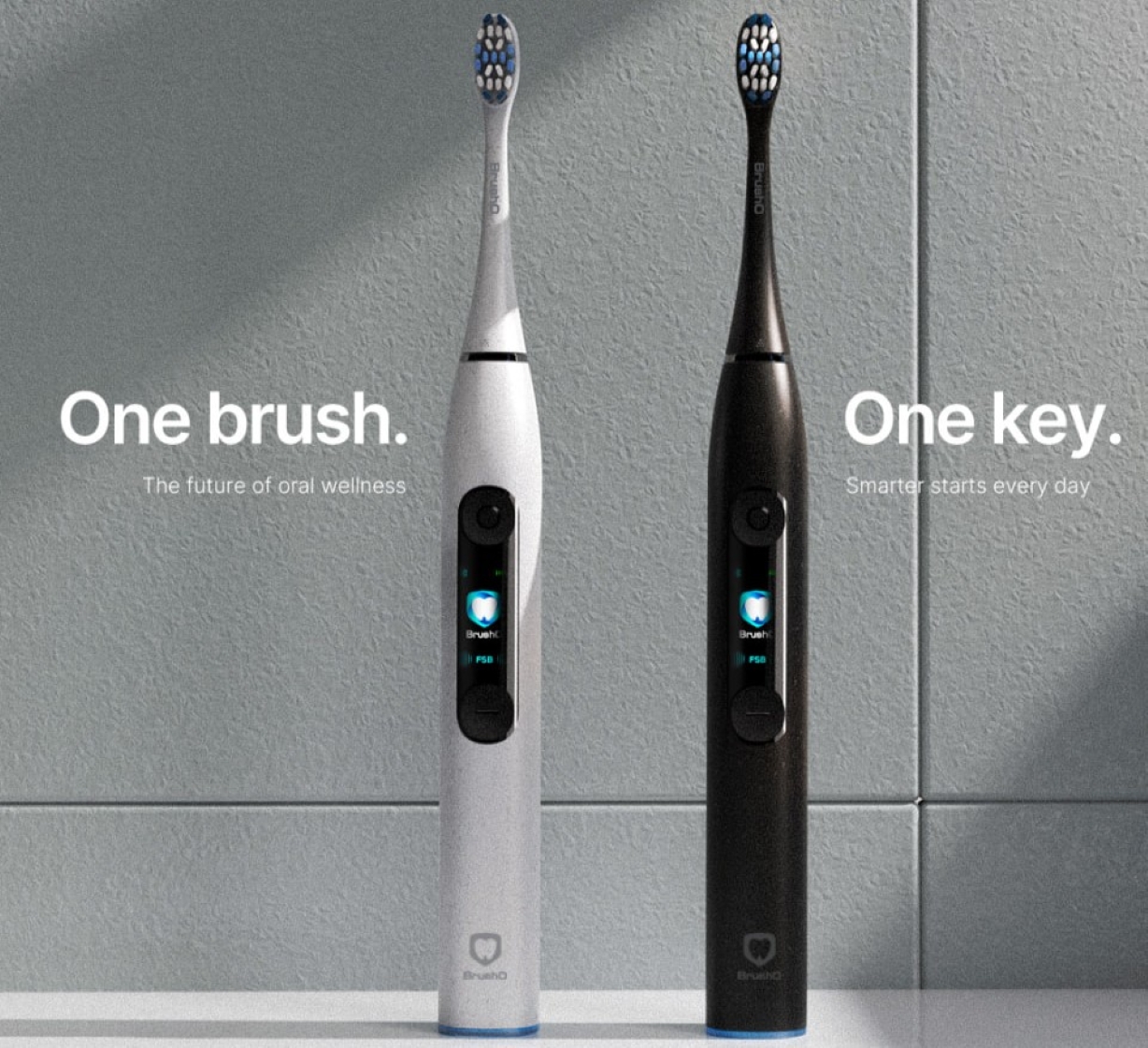
Visible stains aren’t the only sign of wear. Over hundreds of brushing sessions, filaments gradually lose their stiffness, splay outward, and the end-rounded tips flatten—dramatically reducing plaque removal along your gumline and between teeth. Meanwhile, unseen bacteria and food particles can lodge deep within the bristle base.
🪄 BrushO Fix:
Replace your brush head every three to four months regardless of appearance, per ADA guidelines. On BrushO middle and hard heads, a pale-pink wear-indicator stripe fades to signal exactly when to swap.
Topping off your brush after every use may seem safe, but lithium-ion cells actually prefer moderate discharge cycles. Constantly maintaining 100% charge stresses the battery’s chemistry, while allowing it to fully deplete risks deep-discharge damage that can shorten overall lifespan.
🪄 BrushO Fix:
Wait until your battery level drops to around 20% before recharging, then disconnect once full. Plus, run a complete 0→100% cycle every 4–6 weeks to recalibrate the battery management system and preserve long-term capacity.
A damp, enclosed environment is a breeding ground for mold and bacteria—especially in those hard-to-reach bristle bases. Capping or storing your brush head before it fully dries traps moisture, leading to musty odors and potential microbial growth that defeats every brushing effort.
🪄 BrushO Fix:
After rinsing, vigorously shake off excess water, then stand your brush upright in open air until completely dry. Only cap or cover the head once no visible moisture remains.
Relying on feel alone means you miss out on BrushO’s data-driven insights. The app doesn’t just tell you when to replace your head or when to charge; it shows where you’re over- or under-brushing with live coverage heat-maps, logs pressure patterns to protect your gums, and delivers firmware updates that fine-tune brushing modes.
🪄 BrushO Fix:
Sync after every session. Let the app track your pressure, coverage, and wear—then act on its “Head Replacement Recommended” and “Time to Charge” alerts based on real-use metrics, not a calendar.
Neglecting chargers, holders, and any storage cases can silently undermine performance. Dust or toothpaste residue on charging contacts increases resistance and can lead to slow or incomplete charging. Unclean storage stands and travel cases can reintroduce contaminants back onto your brush.
🪄 BrushO Fix:
- Charging Base: Wipe metal contacts weekly with a dry cloth or cotton swab.
- Storage Stands/Travel Cases: Clean monthly with mild soap and warm water; air-dry fully before reassembling.
- Spare Heads: Store extras in a cool, dry place and use color-coded rings to avoid mix-ups and cross-contamination.
Don’t let these myths short-circuit your BrushO’s performance. 🥺
Follow these official care tips—swap heads on schedule, charge smart, dry thoroughly, sync faithfully, and maintain accessories—to unlock the full power of your AI-driven oral-care routine.
Ready to transform your brushing routine?
Discover the full BrushO experience—shop brush heads, download the app, and unlock AI-powered clean at BrushO. Join the smart-brushing revolution today!
Aug 8
Aug 7

Squamous cell carcinoma is not a single, uniform disease, and two of its oral variants illustrate the extremes of its behavior. Verrucous carcinoma is a slow-growing, exophytic tumor that almost never metastasizes, while spindle cell carcinoma is an aggressive, sarcomatoid malignancy that can be ...

Most people expect a toothache to be the first sign of pulp disease, but some forms of pulpitis are surprisingly silent. A pulp polyp is the visible result of chronic hyperplastic pulpitis, a condition in which the dental pulp grows out of a large carious opening as a fleshy mass. Recognizing thi...

Dry mouth is not merely uncomfortable; it is a medical condition with real consequences for teeth, speech, swallowing and quality of life. When the salivary glands cannot produce enough saliva, medications called sialogogues can restore some of the missing fluid. Pilocarpine and cevimeline are th...

A blister in the mouth can be the first warning of a systemic autoimmune disease, and two disorders deserve particular attention in the dental chair: pemphigus vulgaris and mucous membrane pemphigoid. Both produce chronic oral ulceration, yet their immunologic targets, clinical behavior and long-...

When infection drains from a tooth, the body often creates a visible escape route, and the dentist must interpret these fistulous openings correctly. A parulis is the common gum boil that signals chronic periapical infection, while an oroantral fistula is a communication between the mouth and the...

Among the fibro-osseous lesions of the jaws, the ossifying fibroma holds a unique position because it is a true neoplasm, unlike the reactive cemento-osseous dysplasia discussed in an earlier article. It grows continuously, expands bone and requires surgical removal, and its cemental variant adds...

Not all keratocystic lesions of the jaw behave alike, and one of the most important distinctions in oral pathology separates the orthokeratinized odontogenic cyst from its better-known relative, the odontogenic keratocyst. Although the two lesions look similar under the microscope at first glance...

Periodontal disease is usually a slowly progressive infection, but a rare form called necrotizing ulcerative periodontitis destroys tissue with alarming speed. Unlike ordinary periodontitis, this condition produces necrotic, cratered papillae, deep pocketing and rapid bone loss, and it appears al...

Every parent has met the child who refuses to eat because of painful mouth sores, and most of these children have one of two viral illnesses: herpangina or hand, foot and mouth disease. Both are caused by enteroviruses, both produce fever and oral ulcers, and both are overwhelmingly common in you...

Cementoosseous dysplasia is one of the most common fibro-osseous lesions found on dental radiographs, yet it is also one of the most frequently misdiagnosed. The lesion replaces normal bone with cellular fibrous tissue and cementum-like material, and it appears in several forms that range from a ...

Construir rápidamente y a bajo costo una red de datos sobre salud bucal de una manera que todos puedan construir.
Construir rápidamente y a bajo costo una red de datos sobre salud bucal de una manera que todos puedan construir.


Address: RM 1427, 14/F RADIO CITY 505, HENNESSY RD CAUSEWAY BAY, HONG KONG
Code: 000000
Phone: +852-53758652
Email: customerservice@brusho.com