
Gum disease, also known as periodontal disease, is a silent threat that often starts without pain or visible symptoms. However, if left untreated, it can progress to advanced stages, leading to tooth loss and even systemic health complications. Recognizing the earliest signs—such as bleeding gums, bad breath, and subtle inflammation—is crucial for timely treatment and long-term oral health. In this article, we explore the telltale signs of gum disease, what causes it, and how BrushO’s smart brushing system can help you monitor and prevent it. Whether you’ve experienced occasional gum irritation or want to stay ahead of potential problems, understanding the warning signs can empower you to act before it’s too late.
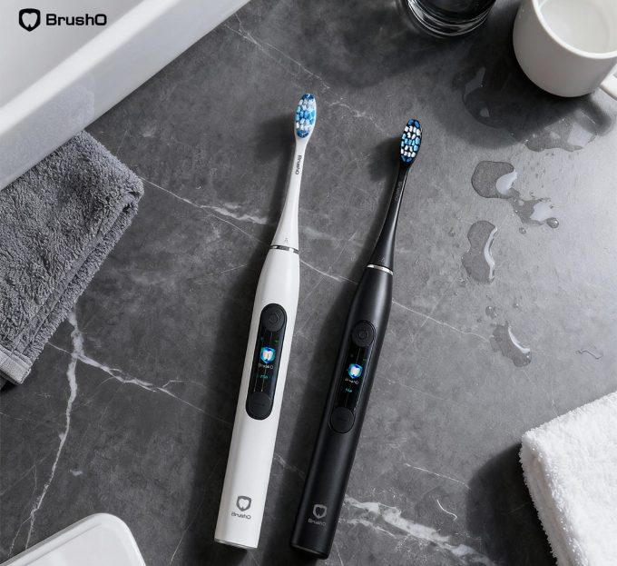
Gum disease typically begins with gingivitis, an early-stage inflammation of the gums caused by plaque buildup. Without intervention, it can progress to periodontitis, which affects the bone that supports your teeth. While early gum disease is reversible, advanced stages may cause irreversible damage.
Bleeding is one of the most obvious and early warning signs. It signals inflammation and irritation from plaque and bacteria near the gum line.
Gum disease causes a buildup of harmful bacteria, releasing foul-smelling compounds. If bad breath lingers despite good hygiene, it could be more than just diet.
Healthy gums are pink and firm. If you notice redness, puffiness, or sensitivity, this may be due to localized inflammation.
Gum recession may indicate tissue damage. If your teeth look longer than before or you feel gaps at the base, your gums may be receding.
As gums pull away and bone support weakens, your teeth may feel more sensitive or even begin to shift.
• Poor oral hygiene
• Skipping nightly brushing
• Using improper brushing techniques
• Smoking or tobacco use
• Genetic predisposition
• Health conditions like diabetes
BrushO’s AI-powered smart toothbrush offers unique features that support gum health monitoring:
• Pressure Sensors: Alerts you when brushing too hard, protecting delicate gum tissue.
• Surface Coverage Analysis: Detects neglected areas near the gum line—where plaque thrives.
• Real-Time Feedback and Reports: Provides reminders and brushing scores to help improve gum care consistency.
• Smart App Integration: Visual charts show brushing coverage and help track problem areas over time.
By improving technique and coverage, BrushO reduces the likelihood of plaque buildup and gum inflammation.
If you experience:
• Consistent gum bleeding
• Gum pain or receding gums
• Wobbly teeth or changes in bite
It’s time to consult a dentist. Early professional intervention can prevent irreversible damage.
• Brush at least twice daily using a pressure-sensitive smart toothbrush like BrushO
• Floss or use interdental brushes daily
• Avoid smoking
• Use fluoride toothpaste
• Visit your dentist for checkups every 6 months
Gum disease doesn’t have to be inevitable. With awareness, the right tools, and timely action, you can detect the early signs before they develop into something serious. BrushO empowers users with real-time feedback and actionable data to take control of their gum health—making every brush a preventive step toward a healthier smile.
Nov 18
Nov 18

Squamous cell carcinoma is not a single, uniform disease, and two of its oral variants illustrate the extremes of its behavior. Verrucous carcinoma is a slow-growing, exophytic tumor that almost never metastasizes, while spindle cell carcinoma is an aggressive, sarcomatoid malignancy that can be ...

Most people expect a toothache to be the first sign of pulp disease, but some forms of pulpitis are surprisingly silent. A pulp polyp is the visible result of chronic hyperplastic pulpitis, a condition in which the dental pulp grows out of a large carious opening as a fleshy mass. Recognizing thi...

Dry mouth is not merely uncomfortable; it is a medical condition with real consequences for teeth, speech, swallowing and quality of life. When the salivary glands cannot produce enough saliva, medications called sialogogues can restore some of the missing fluid. Pilocarpine and cevimeline are th...

A blister in the mouth can be the first warning of a systemic autoimmune disease, and two disorders deserve particular attention in the dental chair: pemphigus vulgaris and mucous membrane pemphigoid. Both produce chronic oral ulceration, yet their immunologic targets, clinical behavior and long-...

When infection drains from a tooth, the body often creates a visible escape route, and the dentist must interpret these fistulous openings correctly. A parulis is the common gum boil that signals chronic periapical infection, while an oroantral fistula is a communication between the mouth and the...

Among the fibro-osseous lesions of the jaws, the ossifying fibroma holds a unique position because it is a true neoplasm, unlike the reactive cemento-osseous dysplasia discussed in an earlier article. It grows continuously, expands bone and requires surgical removal, and its cemental variant adds...

Not all keratocystic lesions of the jaw behave alike, and one of the most important distinctions in oral pathology separates the orthokeratinized odontogenic cyst from its better-known relative, the odontogenic keratocyst. Although the two lesions look similar under the microscope at first glance...

Periodontal disease is usually a slowly progressive infection, but a rare form called necrotizing ulcerative periodontitis destroys tissue with alarming speed. Unlike ordinary periodontitis, this condition produces necrotic, cratered papillae, deep pocketing and rapid bone loss, and it appears al...

Every parent has met the child who refuses to eat because of painful mouth sores, and most of these children have one of two viral illnesses: herpangina or hand, foot and mouth disease. Both are caused by enteroviruses, both produce fever and oral ulcers, and both are overwhelmingly common in you...

Cementoosseous dysplasia is one of the most common fibro-osseous lesions found on dental radiographs, yet it is also one of the most frequently misdiagnosed. The lesion replaces normal bone with cellular fibrous tissue and cementum-like material, and it appears in several forms that range from a ...


Address: RM 1427, 14/F RADIO CITY 505, HENNESSY RD CAUSEWAY BAY, HONG KONG
Code: 000000
Phone: +852-53758652
Email: customerservice@brusho.com
