
Shared bathrooms can be breeding grounds for germs, and your toothbrush might be more exposed than you think. In this blog, we’ll explore the risks of improper toothbrush storage in shared environments and offer smart, science-backed solutions to keep your oral hygiene tools clean and safe. Learn why toothbrush storage matters—and how BrushO makes it easier.
Bathrooms—especially shared ones in dorms, hostels, or family homes—harbor bacteria and moisture that can easily contaminate your toothbrush. Key risks include:
• Aerosol exposure: Flushing toilets release droplets that can land on nearby surfaces—including toothbrushes.
• Cross-contamination: Brushes stored together can touch and transfer bacteria.
• Moisture retention: A wet brush is a perfect breeding ground for mold and bacteria.
Poor storage can undermine even the most careful brushing habits.
Here’s how to protect your brush (and your mouth) in a shared bathroom:
Use a toothbrush holder that keeps each brush standing vertically and spaced apart. Never let brush heads touch.
A ventilated toothbrush cover can protect against splashes, but sealed containers may trap moisture and promote bacterial growth.
Ideally, place your brush at least 1 meter (3 feet) away from the toilet to reduce exposure to toilet plume particles.
Consider a UV sanitizing case if multiple people use the same bathroom. It reduces bacterial load without chemicals.
Always allow your toothbrush to air dry between uses. Bacteria thrive in moist environments.
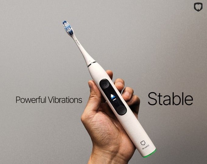
BrushO isn’t just smart when you brush — it’s smart between brushes, too. Features that make it ideal for shared environments:
Each family member can have their own interchangeable brush head with personalized app tracking, avoiding any mix-ups.
See your usage history and replacement reminders — especially helpful if your brush has been exposed or dropped.
Perfect for protecting your toothbrush from shared space exposure when you’re on the go or storing it away safely.
• Label your toothbrush or use color-coded heads.
• Carry your brush in and out of the bathroom using a storage case.
• Don’t share toothpaste — squeeze onto your finger or a clean surface instead.
• Replace your brush head every 2–3 months, or sooner if it’s been contaminated.
In a shared bathroom, proper toothbrush storage is essential. Don’t let invisible germs undo your healthy habits. Combine smart storage habits with BrushO’s smart technology to stay protected—even when bathroom space isn’t private.
BrushO is an AI-powered smart toothbrush that enhances daily oral care with real-time feedback, brushing zone analysis, pressure monitoring, and brushing scores. Whether you’re sharing a bathroom or brushing solo, BrushO helps you maintain a cleaner, safer, and more effective routine.

Squamous cell carcinoma is not a single, uniform disease, and two of its oral variants illustrate the extremes of its behavior. Verrucous carcinoma is a slow-growing, exophytic tumor that almost never metastasizes, while spindle cell carcinoma is an aggressive, sarcomatoid malignancy that can be ...

Most people expect a toothache to be the first sign of pulp disease, but some forms of pulpitis are surprisingly silent. A pulp polyp is the visible result of chronic hyperplastic pulpitis, a condition in which the dental pulp grows out of a large carious opening as a fleshy mass. Recognizing thi...

Dry mouth is not merely uncomfortable; it is a medical condition with real consequences for teeth, speech, swallowing and quality of life. When the salivary glands cannot produce enough saliva, medications called sialogogues can restore some of the missing fluid. Pilocarpine and cevimeline are th...

A blister in the mouth can be the first warning of a systemic autoimmune disease, and two disorders deserve particular attention in the dental chair: pemphigus vulgaris and mucous membrane pemphigoid. Both produce chronic oral ulceration, yet their immunologic targets, clinical behavior and long-...

When infection drains from a tooth, the body often creates a visible escape route, and the dentist must interpret these fistulous openings correctly. A parulis is the common gum boil that signals chronic periapical infection, while an oroantral fistula is a communication between the mouth and the...

Among the fibro-osseous lesions of the jaws, the ossifying fibroma holds a unique position because it is a true neoplasm, unlike the reactive cemento-osseous dysplasia discussed in an earlier article. It grows continuously, expands bone and requires surgical removal, and its cemental variant adds...

Not all keratocystic lesions of the jaw behave alike, and one of the most important distinctions in oral pathology separates the orthokeratinized odontogenic cyst from its better-known relative, the odontogenic keratocyst. Although the two lesions look similar under the microscope at first glance...

Periodontal disease is usually a slowly progressive infection, but a rare form called necrotizing ulcerative periodontitis destroys tissue with alarming speed. Unlike ordinary periodontitis, this condition produces necrotic, cratered papillae, deep pocketing and rapid bone loss, and it appears al...

Every parent has met the child who refuses to eat because of painful mouth sores, and most of these children have one of two viral illnesses: herpangina or hand, foot and mouth disease. Both are caused by enteroviruses, both produce fever and oral ulcers, and both are overwhelmingly common in you...

Cementoosseous dysplasia is one of the most common fibro-osseous lesions found on dental radiographs, yet it is also one of the most frequently misdiagnosed. The lesion replaces normal bone with cellular fibrous tissue and cementum-like material, and it appears in several forms that range from a ...


Address: RM 1427, 14/F RADIO CITY 505, HENNESSY RD CAUSEWAY BAY, HONG KONG
Code: 000000
Phone: +852-53758652
Email: customerservice@brusho.com
