
Many people unconsciously focus only on the visible front surfaces of their teeth when brushing — especially during rushed mornings or late-night routines. But what happens to the areas you skip? Neglecting the back sides, molars, and inner surfaces can lead to plaque buildup, gum inflammation, and serious oral health issues over time. In this article, we explore why brushing only the front of your teeth isn’t enough and how smart toothbrushes like BrushO help you achieve thorough, all-around cleaning every time.
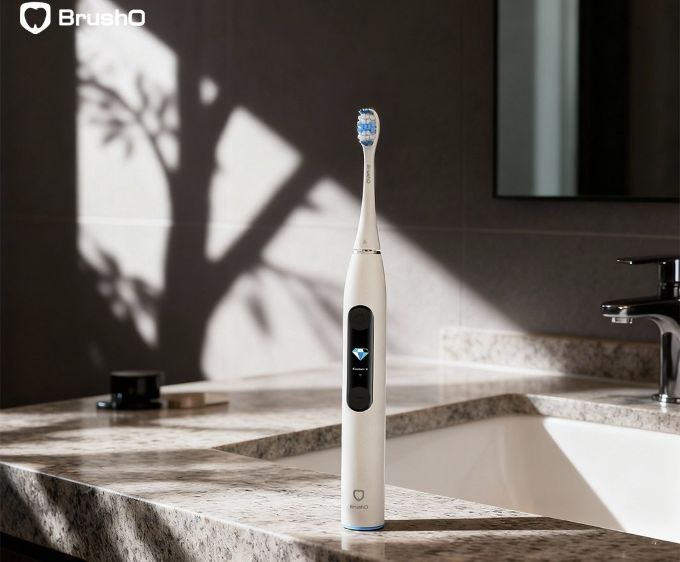
It’s common to brush what you can easily see in the mirror — mostly the front-facing surfaces. But behind every smile lies a complex set of surfaces, including:
• Back of front teeth
• Inner molars
• Gumline areas
• Chewing surfaces
Many people unintentionally rush through these hidden zones, leaving behind food particles and bacteria.
Plaque forms on all surfaces of your teeth, not just the front. When you skip the back or inner surfaces, plaque and bacteria can grow unchecked — especially in hard-to-reach molars and along the gumline.
Cavities often form between teeth or on the back surfaces where brushing is less thorough. Focusing only on the front makes you more likely to miss areas where food debris hides.
Gum disease starts where plaque isn’t removed — typically near the back molars and inner gumline. Inflammation, bleeding, and even recession can follow.
Bacteria lingering on back teeth or the back of your tongue can cause persistent bad breath, even if your front teeth look spotless.
Brushing thoroughly — front, back, sides, and chewing surfaces — is essential. BrushO is designed to make this easy, effective, and even automatic.
BrushO uses AI-powered sensors and algorithms to monitor which zones you’ve cleaned and which ones you’ve missed. If the back molars or inner teeth haven’t been brushed adequately, the app will notify you in real time.
Unlike traditional electric toothbrushes that use a 4-quadrant timer, BrushO divides the mouth into 6 zones and 16 surfaces, giving precise coverage for:
• Front and back of each tooth
• Upper and lower jaws
• Inner and outer surfaces
This ensures no area is skipped.
Each brushing session is scored based on coverage, pressure, and duration. Users see exactly which areas need more attention and improve their habits over time.
The BrushO app generates heatmaps to visualize brushing coverage. You’ll know at a glance if you’re favoring the front and missing the back — and learn how to correct it.
• Angle the brush: Tilt your brush at a 45° angle to clean the gumline effectively.
• Use a mirror: Occasionally check to ensure you’re reaching the back teeth.
• Don’t rush: Spend at least 2 minutes brushing, 30 seconds per major section.
• Let the toothbrush do the work: With smart devices like BrushO, you only need to guide — the AI handles the precision.
Only brushing the front of your teeth may leave you with a nice-looking smile in the mirror, but it’s what happens in the areas you can’t see that truly determines your oral health. By brushing every surface of every tooth — and getting a little help from BrushO’s smart tech — you can prevent decay, reduce gum disease risk, and maintain a truly healthy mouth.

Squamous cell carcinoma is not a single, uniform disease, and two of its oral variants illustrate the extremes of its behavior. Verrucous carcinoma is a slow-growing, exophytic tumor that almost never metastasizes, while spindle cell carcinoma is an aggressive, sarcomatoid malignancy that can be ...

Most people expect a toothache to be the first sign of pulp disease, but some forms of pulpitis are surprisingly silent. A pulp polyp is the visible result of chronic hyperplastic pulpitis, a condition in which the dental pulp grows out of a large carious opening as a fleshy mass. Recognizing thi...

Dry mouth is not merely uncomfortable; it is a medical condition with real consequences for teeth, speech, swallowing and quality of life. When the salivary glands cannot produce enough saliva, medications called sialogogues can restore some of the missing fluid. Pilocarpine and cevimeline are th...

A blister in the mouth can be the first warning of a systemic autoimmune disease, and two disorders deserve particular attention in the dental chair: pemphigus vulgaris and mucous membrane pemphigoid. Both produce chronic oral ulceration, yet their immunologic targets, clinical behavior and long-...

When infection drains from a tooth, the body often creates a visible escape route, and the dentist must interpret these fistulous openings correctly. A parulis is the common gum boil that signals chronic periapical infection, while an oroantral fistula is a communication between the mouth and the...

Among the fibro-osseous lesions of the jaws, the ossifying fibroma holds a unique position because it is a true neoplasm, unlike the reactive cemento-osseous dysplasia discussed in an earlier article. It grows continuously, expands bone and requires surgical removal, and its cemental variant adds...

Not all keratocystic lesions of the jaw behave alike, and one of the most important distinctions in oral pathology separates the orthokeratinized odontogenic cyst from its better-known relative, the odontogenic keratocyst. Although the two lesions look similar under the microscope at first glance...

Periodontal disease is usually a slowly progressive infection, but a rare form called necrotizing ulcerative periodontitis destroys tissue with alarming speed. Unlike ordinary periodontitis, this condition produces necrotic, cratered papillae, deep pocketing and rapid bone loss, and it appears al...

Every parent has met the child who refuses to eat because of painful mouth sores, and most of these children have one of two viral illnesses: herpangina or hand, foot and mouth disease. Both are caused by enteroviruses, both produce fever and oral ulcers, and both are overwhelmingly common in you...

Cementoosseous dysplasia is one of the most common fibro-osseous lesions found on dental radiographs, yet it is also one of the most frequently misdiagnosed. The lesion replaces normal bone with cellular fibrous tissue and cementum-like material, and it appears in several forms that range from a ...

সবাই যেন নিজের মতো করে গড়তে পারে – এমনভাবে দ্রুত ও কম খরচে ওরাল হেলথ ডেটা নেটওয়ার্ক তৈরি করুন।
সবাই যেন নিজের মতো করে গড়তে পারে – এমনভাবে দ্রুত ও কম খরচে ওরাল হেলথ ডেটা নেটওয়ার্ক তৈরি করুন।


Address: RM 1427, 14/F RADIO CITY 505, HENNESSY RD CAUSEWAY BAY, HONG KONG
Code: 000000
Phone: +852-53758652
Email: customerservice@brusho.com